Something to ponder.

For those who fear that acknowledging the historical fact of evolution dooms one to a life of bleak insignificance, consider the following.

You are the product of an absolutely unbroken chain of successful ancestors stretching back nearly 4 billion years. In all that time, over billions of generations, not one member of your lineage ever failed to leave viable offspring who, in their turn, left yet more successful descendants. Not a single earthquake, volcanic eruption, meteorite impact, or glacier ever prevented one of your ancestors from contributing to the subsequent generation. Every one of your forebears prevailed in the face of predators, famines, parasites, diseases, and ill fortune. Whether in competition or cooperation, your antecedents triumphed. An untold number of beings have lived and died on this planet, but never — not a single time — did your line falter.

In this, you are not alone. The same is true of every living being on Earth, to whom you are connected directly through converging lines of common ancestry that date back to the very dawn of life. The world did not know you were coming and the machinations of nature did not have you in mind as an endproduct, yet here you are. As an individual, you and each of your brethren, cousins, and more distant evolutionary relatives represent an exceedingly, remarkably, staggeringly improbable occurrence — and are all the more wonderful for it.


From "Pangenesis" to "Genome".

The term “genetics” has been used in reference to the branch of science dealing with “the physiology of heredity and variation” since 1905. It was coined by the British biologist William Bateson, first in a 1905 letter (see Bateson 1928), and then publicly the following year (Bateson 1906). It was derived directly from the Greek for “birth” (or “origins”).

Straightforward enough. But what about “gene” and “genome”? These terms are interesting because they illustrate the evolution of both concept and language in science and involve both co-option and hybridization.

First, “gene”. Even after the term “genetics” was in use, it was not entirely clear what practitioners of the science were studying. Indeed, the concept of a fundamental physical and functional unit (or “determiner”) of heredity remained very vague. In 1909, Danish biologist Wilhelm Johannsen sought to pin down a term to describe these genetic elements. Although some people attribute the origin of “gene” to the same etymology as “genetics”, there is more to the story. In actuality, “gene” was derived indirectly from Darwin‘s (incorrect) theory of heredity known as “pangenesis“. Indirectly, because it morphed through the term “pangens” coined by the Dutch botanist Hugo de Vries in 1889 in reference to genetic units and as an homage to Darwin, even though his theory of heredity differed markedly from pangenesis (de Vries was a Mendelian).

According to Johannsen (1909, p.143), he came up with the term “gene” by choosing to isolate

the last syllable ‘gene’, which alone is of interest to us, from Darwin’s well known word (Pangenesis) and thereby replace the less desirable ambiguous word ‘determiner’. Consequently, we will speak of ‘the gene’ and ‘the genes’ instead of ‘pangen’ and ‘the pangens’. The word gene is completely free from any hypothesis; it expresses only the evident fact that, in any case, many characteristics of the organism are specified in the germ cells by means of special conditions, foundations, and determiners which are present in unique, separate, and thereby independent ways – in short, precisely what we wish to call genes. [Translation as in Portugal and Cohen 1977].

Johannsen (1909) was also responsible for the terms “genotype” and “phenotype“. As he summarized in 1911,

I have proposed the terms ‘gene’ and ‘genotype’ … to be used in the science of genetics. The ‘gene’ is nothing but a very applicable little word, easily combined with others, and hence it may be useful as an expression for the ‘unit-factors’, ‘elements’ or ‘allelomorphs’ in the gametes, demonstrated by modern Mendelian researches. A ‘genotype’ is the sum total of all the ‘genes’ in a gamete or in a zygote.

So, we have an evolution of the term from “pangenesis” (Darwin) to “pangens” (de Vries) to “genes” (Johannsen), passing through an incorrect theory of heredity to a term “completely free from any hypothesis” about inheritance to Mendelian genetics.

What about “genome”?

According to the Oxford English Dictionary, the term “genom(e)” was coined by the German botanist Hans Winkler in 1920 as a portmanteau of gene and chromosome (the latter term having been coined by Wilhelm Waldeyer in 1888). This story has been repeated by many authors (including yours truly; Gregory 2001), but has been challenged by Lederberg and McCray (2001), who suggest that Winkler probably merged gene with the generalized suffix ‘ome (referring to “the entire collectivity of units”), and not ‘some (“body”) from chromosome. In either case, Winkler’s intent was to “propose the expression Genom for the haploid chromosome set, which, together with the pertinent protoplasm, specifies the material foundations of the species” (translation as in Lederberg and McCray 2001).

Based on this initial formulation, “genome” can accurately be taken to mean either the total gene complement (interchangeably with Johannsen’s “genotype”), or the total DNA amount per haploid chromosome set – but not both, as we now know that these are not correlated with one another. This latter issue remains the subject of active study, and I shall have much more to say about it in future postings.

__________

References

Bateson, W. 1906. A text-book of genetics. Nature 74: 146-147.

Bateson, W. 1928. Letter to Sedgwick, April 18, 1905. In William Bateson, F.R.S.: His Essays and Addresses (ed. B. Bateson), pp. 93. Cambridge University Press, Cambridge.

De Vries, H. 1889. Intrazelluläre Pangenesis. Fischer, Jena.

Gregory, T.R. 2001. The bigger the C-value, the larger the cell: genome size and red blood cell size in vertebrates. Blood Cells, Molecules, and Diseases 27: 830-843.

Johannsen, W. 1909. Elemente der Exakten Erblichkeitslehre. Fischer, Jena.

Johannsen, W. 1911. The genotype conception of heredity. American Naturalist 45: 129-159.

Lederberg, J. and A.T. McCray. 2001. ‘Ome sweet ‘omics — a genealogical treasury of words. The Scientist 15: 8.

Portugal, F.H. and J.S. Cohen. 1977. A Century of DNA. MIT Press, Cambridge, MA.

Winkler, H. 1920. Verbeitung und Ursache der Parthenogenesis im Pflanzen und Tierreiche. Verlag Fischer, Jena.


Chimps are not more evolved than humans or anyone else.

I like New Scientist. I even did a short interview with them about a cool genomics story (“How chemicals can speed up evolution“, 6 May 2006, p.16). But this headline from their news service really annoys me: Chimps ‘more evolved’ than humans.

The short news article starts out with “It is time to stop thinking we are the pinnacle of evolutionary success…”, which of course is true except that it was time to stop thinking this 150 years ago, and then continues with “… chimpanzees are the more highly evolved species, according to new research”.

What they mean is that, based on the recent study, it appears that the rate of fixation by selection of mutations apparently has been higher in the lineage that has led to chimpanzees than in the lineage that has led to humans since they split from a common ancestor several million years ago. Which lineage experienced the changes can now be inferred by comparison with the macaque genome, which is less closely related to chimps and humans than the latter two are to each other; without such an external comparison, one can not say which lineage had changed, only that one or both of them had. Most likely, this boils down to differences in long-term historical population sizes in the two lineages (selection is stronger in large populations, genetic drift in small populations).

Couching this interesting finding in terms of who is “more evolved” than whom is not helpful, even with the scare quotes. As someone who teaches evolution at the upper-year undergraduate level, I can tell you that students come into the class with a lot of preconceptions about evolution, one of them being the notion that some extant species can be ranked as “more evolved” than others. It is subtle misinformation like this, compounded over many years, that makes my job harder by the time they arrive in my course.

Please, please, PLEASE stop appealing to common misconceptions about evolution in news stories, even if the headline will catch the attention of (previously misinformed) readers.

_________

Updates:


Genome sequences reduce the complexity of bacterial flagella.

I am not interested in engaging in debates with anti-evolutionists, though I am well aware of their key arguments. The big one, of course, is “irreducible complexity” — traits or features that supposedly could not have evolved because there is no conceivable function for their parts individually nor for a subset of their parts collectively. The bacterial flagellum apparently is the ultimate example of this, which explains why this microscopic protein “motor” can drive an entire philosophical argument along these lines.

I think Darwin said it best (as he often did) in 1871: “Ignorance more frequently begets confidence than does knowledge; it is those who know little, and not those who know much, who so positively assert that this or that problem will never be solved by science.”

There is little concern among biologists that the evolution of bacterial flagella will be worked out, just as a tremendous amount of information is now available about the evolution of eyes (the previous Paleyan example of a supposedly un-evolvable structure).

Last year, Pallen and Matzke (2006) presented a discussion of how bacterial flagella may have evolved, based in large part on comparisons of sequences from the various protein components. Many of the proteins that make up a flagellum have homologues that serve non-flagellar functions, strongly suggesting that they were co-opted from pre-existing proteins during the evolution of flagella. (See Matzke’s detailed model of flagellar evolution here and a video based on it here, and Ken Miller talking about flagella here). Specifically, there is ever-mounting evidence that bacterial flagella and the type III secretory system (TTSS) that toxic bacteria use to inject their prey are descended from the same ancestral structure. The fact that the TTSS lacks many of the proteins in flagella but remains functional (for toxin injection rather than locomotion) clearly indicates that not all the parts need to be present for some function to be carried out by the structure.

Pallen and Matzke (2006) noted that further comparisons of complete genome sequences (hence the post on this blog) would reveal additional insights into the evolution of flagella. Enter Liu and Ochman (2007) from the next issue of PNAS.

Liu and Ochman (2007) examined complete genome sequences from 41 species of bacteria with flagella, and were able to identify a core set of 24 proteins common to all of them, which was present in a very early ancestral bacterium. Not only this, but the core genes appear to be the product of multiple rounds of duplication and diversification, perhaps of one original precursor gene.

The gist of the story is that 1) some genes involved in the construction of flagella in modern bacteria are clearly co-opted from pre-existing genes that were doing something else in the cell (Pallen and Matzke 2006) and 2) a core of about two dozen genes common to all flagellated bacteria (and presumably found in their common ancestor) is the product of duplication and divergence whose reconstructed history agrees very well with the presumed evolutionary relationships among bacteria (Liu and Ochman 2007).

This just goes to show the usefulness of genome data for addressing questions that, for the reason outlined by Darwin, seem unanswerable to some. It also opens the door to some exciting future work.

I asked Howard Ochman what he thought the next key steps will be in this line of study. As he put it, “Naturally we would like to know the function of the structures that were specified by the ancestral set of flagellar genes, and how/why these genes remained functional through their successive duplications. We just completed a companion paper on the bacterial flagellar genes that arose later, and we are now branching out in into the other domains of life.”

I will positively assert, out of optimism rather than ignorance, that many more important insights will be forthcoming from these investigations.

(Update: Nick Matzke is very critical of the paper. He also has posted an updated critique that focuses more on the data.)

(Another update: See Carl Zimmer’s post about blogging as scientific debate).

(And yet another update: A complex tail, simply told at ScienceNOW)

_________

References

Aizawa, S.-I. 2001. Bacterial flagella and type III secretion systems. FEMS Microbiology Letters 202: 157-164.

Blocker, A., K. Komoriya, and S.-I. Aizawa. 2003. Type III secretion systems and bacterial flagella: insights into their function from structural similarities. Proceedings of the National Academy of Sciences of the USA 100: 3027-3030.

Gophna, U. , E.Z. Ron, and D. Graur. 2003. Bacterial type III secretion systems are ancient and evolved by multiple horizontal-transfer events. Gene 312: 151–163.

Liu, R. and H. Ochman. 2007. Stepwise formation of the bacterial flagellar system. Proceedings of the National Academy of Sciences of the USA 104: 7116-7121.

Matzke, N.J. 2003. Evolution in (Brownian) space: a model for the origin of the bacterial flagellum. Talk.Origins.

Miller, K.R. 2004. The flagellum unspun. In Debating Design: From Darwin to DNA, edited by W. Dembski and M. Ruse. Cambridge University Press, New York, pp. 81-97.
(available online here)

Musgrave, Ian. 2004. Evolution of the bacterial flagellum. In Why Intelligent Design Fails: A Scientific Critique of the New Creationism, edited by M. Young and T. Edis. Rutgers University Press, New Brunswick, NJ.
(available online here)

Nguyen, L., I.T. Paulsen, J. Tchieu, C.J. Hueck, and M.H. Saier. 2000. Phylogenetic analyses of the constituents of Type III protein secretion systems. Journal of Molecular Microbiology and Biotechnology 2: 125–144.

Pallen, M.J., C.W. Penn, and R.R. Chaudhuri. 2005. Bacterial flagellar diversity in the post-genomic era. Trends in Microbiology 13: 143-149.

Pallen, M. J., S.A. Beatson, and C.M. Bailey. 2005. Bioinformatics, genomics and evolution of non-flagellar type-III secretion systems: a Darwinian perspective. FEMS Microbiology Reviews 29: 201–229.

Pallen, M.J. and N.J. Matzke. 2006. From The Origin of Species to the origin of bacterial flagella. Nature Reviews Microbiology 4: 784-790.


DNA barcoding and taxonomy funding.

This may be old news, but it seems worthwhile responding anyway because hey, I have a blog now.

In 2005, Ebach and Holdrege wrote a letter to Nature in which they repeated the common misconception that DNA barcoding steals funding from taxonomic research. I responded by pointing out that DNA barcoding has not drawn support from the taxonomy pool, but rather so far has competed with medicine and genomics for support, or has brought in funds from sources that have not traditionally supported taxonomy. Apparently John Wilkins of Evolving Thoughts was not buying it.


I feel somewhat qualified to speak on this because I co-authored a Genome Canada grant with Paul Hebert that helped to fund the Canadian Barcode of Life Network. Needless to say, Genome Canada does not normally fund taxonomy. Other sources of support have been the Moore Foundation, the Sloan Foundation, the Canada Foundation for Innovation, NSERC, and various other government and industry sources — none of which has been taken from taxonomists (and in fact, a lot of it goes directly to taxonomists).

The “barcoders versus taxonomists” dichotomy, which is taken as a given by Ebach and Holdrege and other opponents of barcoding, is false. DNA barcoding is a collaborative enterprise, requiring people with different expertise. Existing DNA barcoding networks involve a large number of professional taxonomists, and major participants include museums and other taxonomic institutions. It also is a gross caricature to suggest that those who come at the issue from a molecular perspective are not interested in organismal biology. Paul Hebert, considered the father of DNA barcoding, spent most of his career cataloguing the diversity and phylogeography of aquatic microcrustaceans. This included both morphological and molecular work and even extended to development of traditional taxonomic keys. Many of the rest of us are proponents of barcoding because it allows us to access information about organisms that otherwise is inaccessible to non-taxonomists.

As far as I know, Wilkins is not a biologist, which means that his work is not contingent on being able to obtain species identifications. The situation is very different for biodiversity researchers. I have plenty of genome size estimates that remain unpublished because, try as I might, I cannot get them identified. Even assuming that a suitable taxonomic expert exists, he or she may be backlogged by a year or more. This is why DNA barcoding proponents consider it an enabling technology — a lot more scientists need taxonomy than do taxonomy.

As Wilkins notes,
“In computing terms, a DNA barcode is at best a record ID. The details of the record – the rest of the fields describing the name, address, and personal details of the species – still need to be recorded. Otherwise, all you have are empty records…”

I personally don’t know any DNA barcoders who would disagree with this. DNA barcoding is not the endpoint of biodiversity research, it is an access point. It may help to identify “groups” of genetically similar organisms, but the description of those groups, if warranted, will be done according to traditional taxonomic principles. This would not even be an issue, and DNA barcoding could focus only on its primary objective of identification, if not for the fact that most of life has yet to be described. If anything, barcoding should make these new taxonomic descriptions both easier and more accessible, which is the reason that so many taxonomists are working as part of the DNA barcoding initiative.

DNA barcoders are interested in information about biodiversity, and they want it to be accessible to everyone — and so far, they have worked to accomplish this without siphoning funds from the existing taxonomic pot.

__________

References

Ebach, M.C. and C. Holdrege. 2005. DNA barcoding is no substitute for taxonomy. Nature 434: 697.

Gregory, T.R. 2005. DNA barcoding does not compete with taxonomy. Nature 434: 1067.

Hebert, P.D.N. and T.R. Gregory. 2005. The promise of DNA barcoding for taxonomy. Systematic Biology 54: 852-859.
(Check out the cover)

Schindel, D.E. and S.E. Miller. 2005. DNA barcoding a useful tool for taxonomists. Nature 435: 17.

Links


Whose genome?

The term “genome” is oft-heard but seldom defined, and indeed has more than one meaning. Little wonder, then, that discussions about genome sequences and comparisons thereof can leave otherwise interested audiences more frustrated than enlightened. “What is a genome?” and “whose genome was sequenced?” are legitimate questions, and what follows is an attempt at clarification that is, by necessity, as much philosophical as scientific.

Definition #1: In a broad sense, a genome can be considered as the collective set of genes, non-coding DNA sequences, and all their variants that are located within the chromosomes of members of a given species. This definition does not consider variation among individuals within a species, and instead relates to distinctions between species. It is possible to apply such a definition because, for the most part, animal species do not share DNA extensively and hence their respective gene pools remain distinct (in fact, this forms the basis for defining species under some views). Thus, even though humans and chimpanzees are about 98% identical in terms of their DNA sequences, there is still such as thing as a “human genome” and a “chimpanzee genome” rather than a continuum with humans and chimps at two mildly divergent extremes. This is even true of far closer (but now extinct) relatives of humans such as Neanderthals; on average, the sections of Neanderthal DNA that have been recovered and sequenced are 99.5% identical to that of humans — but these, too, are considered to be part of a separate genome.

The genomic similarities described between species are usually based on comparing a few specific regions of DNA from a small number of representative individuals. If other factors are included in the comparison, such as insertions and deletions of DNA, then any two genomes will register a lower level of similarity — say, 95% for chimpanzees and humans rather than 98%. And indeed, no one would ever mistake a chimpanzee genome for a human genome, in part because they differ in DNA amount and chromosome number (human chromosome 2 is a product of fusion of what remain as two separate chromosomes in other great apes).

Of course, individuals within species are not genetically identical to one another (monozygotic twins notwithstanding), which leads to definition #2.

Definition #2: Because the DNA sequences of even close family members are not identical, it can also be said that each individual carries a unique genome consisting of the DNA in his or her chromosomes. In this case, the focus is entirely on one species and the important factor is the variability that exists among individuals of that species. In terms of DNA sequences on a large scale, members of the same species are extremely similar: overall, any two human beings are probably about 99.9% the same genetically. Nevertheless, complete genome sequencing, though conducted primarily under definition #1, has revealed two major sources of variation among individuals. The first are known as single nucleotide polymorphisms (SNPs, “snips”) and are as their name implies: differences at the level of single base pairs that are present in at least 1% of the population. It is estimated that there are some 3 million SNPs in the human genome (definition #1), with one occurring about every 100-300 base pairs along the more than 3 billion base pair sequence. The second major source of variation, first described in 2006, are known as copy number variants (CNVs). These involve differences among individuals in the insertion and deletion of larger DNA segments. CNVs have proved to be far more common than anyone would have imagined, and can result in differences not just in sequences but in the sizes of genomes among individuals (up to 20 million base pairs in humans, or about 0.5%).

The human genome, definition #1

Two independent research groups reported draft sequences of the human genome in February 2001: the publicly-funded and internationally collaborative Human Genome Project and the private company headed by J. Craig Venter known as Celera Genomics. The interaction of these two initiatives – typically branded as competitive, but also mutually informative – has been discussed many times. The question of how and why the two groups sequenced human DNA is not the subject of interest here – the question at present is whose DNA they analyzed.

The Human Genome Project, being a public effort, had an official policy of releasing all sequence data to public databases within 24 hours of completion, thereby making the information freely available to anyone who carried a copy of the “human genome” in their cells. In keeping with this outlook, the HGP implemented procedures intended to circumvent the focus on individuality and to keep their results in line with definition #1. Thus, they instituted a policy of voluntary donation by dozens of men and women from various ethnic backgrounds, provided samples with random numeric labels, shipped the samples to processing laboratories where they were re-labeled with new randomized codes, destroyed all records of previous labels, and then selected randomly from among the samples. Five to ten samples were collected for every one that was actually assayed, with the source of samples used unknown to both researchers and donors. In other words, the intent was to focus on definition #1 as much as possible and to provide a mixture, or at least a mystery, when it came to the source of the genome sequence of Homo sapiens.

Human nature being what it is, it is likely that most people would find this answer disappointing. Deep down, we want to know whose genome it was. The only information that has been available in this regard is that the largest portion of the source DNA came from a male donor in Buffalo, New York, code named “RPCI-11” (for Roswell Park Cancer Institute, where the genomic library was generated). No name, no other information, and yet somehow it seems satisfying to know that there really is an individual human – a real person in a specific part of the world who walked into a lab, stuck out his arm, and donated his blood – corresponding to all those A’s, T’s, G’s, and C’s.

The situation at Celera was quite different in terms of both data sharing and DNA sampling policies. Celera’s data were not made publicly available during the course of sequencing, and their sampling involved 20 donors, five of which were selected for analysis — though evidently not entirely at random. In fact, it was later revealed that Celera’s president and lead investigator, J. Craig Venter, was the primary source of DNA for the sequence. Venter argued that revealing this fact would dispel the myth of a single “human genome” (i.e., an excessive emphasis on definition #1 that ignores the individual uniqueness inherent in definition #2). Others may have felt that sequencing his own genome made the resulting sequence the property of one individual rather than of humanity at large (i.e., adopting definition #2 exclusively at the expense of the broadly shared definition #1).

The human genome, definition #2

While the Human Genome Project and Celera’s efforts generated single (partially composite) genome sequences, another major initiative is underway which focuses on variation among individuals at the genomic level; i.e., on definition #2. The International HapMap Project aims to identify associated collections of SNPs known as haplotypes, and currently includes samples from 270 people drawn from four major groups. Thirty sets of “trios” (two parents and a child) have come from the Yoruba people of Ibadan, Nigeria. Forty-five unrelated individuals from Tokyo and 45 from Beijing have provided samples. Thirty trios from residents of the United States with roots in western and northern Europe have also been included. SNP haplotypes may vary among populations and are important in the search for particular genes of medical significance. As with the DNA sequence information of the Human Genome Project, data from the HapMap Project are made freely available. A similar initiative to catalogue human diversity from the perspective of CNVs, the Copy Number Variation Project, has also been launched.

“Whose genome?” and individual identity

The question “whose genome was sequenced?” is predicated on concepts of individuality and personhood, which tend to be applied to the members of only a handful of species. Thus, one might be interested in which strain of fruit fly (Drosophila melanogaster), which population of sea urchins (Strongylocentrotus purpuratus), or which varieties of rice (Oryza sativa) had been sequenced, but it would not make sense to ask “who” the fly, sea urchin, or rice plant was. The situation gains complexity when dealing with vertebrate genomes because humans associate closely and emotionally with members of some species and not with others. The desire (or not) to know “who” was sequenced correlates directly with this. By way of example, consider the fact that a single male pufferfish (Takifugu rubripes), a single female chicken (red jungle fowl, Gallus gallus), two female brown rats (Rattus norvegicus), and a small number of female mice (Mus musculus) of the B6 strain have been sequenced, but that there has not been much interest in “who” these individuals were – nor would many people even think to ask the question.

Now consider “man’s best friend”. Not only is it known that the two reported canine genome sequences were from individual dogs, but it is known who those dogs were: Craig Venter’s poodle, Shadow, and a boxer named Tasha. It was also widely noted that samples for the chimpanzee genome were taken from a captive-born male named Clint who lived at the Yerkes National Primate Research Center in Atlanta, Georgia. Indeed, many a news story reported Clint’s untimely death in 2005 at the young age of 24. One might be tempted to argue that intelligence is the determining factor in this case – dogs and chimps are smart and have personalities, but pufferfish and rats do not. Perhaps. But surely the recently sequenced rhesus macaque (whatever her name was) should qualify under these criteria.

In the end, this post is not meant to be a statement about the apparent arbitrariness of our decisions to grant or deny individuality to members of other species. This is about genomes, and how definition #1 is applied intuitively and automatically when dealing with a species like mouse or rat, but that one cannot help but invoke definition #2 when dealing with a dog or human. The fact is that all of these species are composed of variable individuals, each with a unique genome under definition #2. Indeed, it is this variation that makes evolutionary divergence – and thus definition #1 – possible at all.


Yet another link between dinosaurs and birds.

Score another point for Darwin’s bulldog. T.H. Huxley, who argued that birds are descended from dinosaurs nearly 140 years ago (Huxley 1868), would undoubtedly be pleased with himself were he privy to the wealth of data that has accrued in support of his conjecture. Evidence from the rather good fossil records of dinosaurs and birds strongly supports this notion, and now two additional lines of evidence can be added in its favour.

The first relates to my own area of study, that of genome size diversity. In a recent paper in Nature, Organ et al. used the link between genome size and cell size of extant taxa (in this case, osteocyte spaces) to infer genome sizes for extinct dinosaurs from various lineages (see Zimmer 2007 f0r a helpful overview). I have promised to post a detailed discussion of the genome size-flight issue another time, so I will briefly note that the main finding is that saurischian dinosaurs, the lineage from within which birds are thought to be descended, had small genomes relative to other reptiles, including ornithischian dinosaurs. This suggests that genome size reduction began (but probably did not culminate) prior to the origin of avian flight and provides yet another intriguing link between saurischian dinosaurs and birds.

The second was reported in papers published by Asara et al. and Schweitzer et al. in the April 13 issue of Science, and revealed intriguing similarities in the amino acid sequences of collagen proteins from a 68 million year old bone from Tyrannosaurus rex and that of a modern chicken. Specifically, amino acid sequence identity was closer to chicken (and presumably to all other modern birds) (58%) than to frog or newt (51%). Note that collagen sequence is usually quite conserved and that what the authors were dealing with were fragments of proteins. Note also that some other interesting comparisons — especially with other living archosaurs, namely alligators or crocodiles — were not possible based on the currently available data. Plenty more to do as follow up to this study, but a very interesting result. A summary of the studies can be found in National Geographic and the New York Times.

This is a nice example of the way in which independent sources of information converge on a common conclusion in evolutionary biology, and how new discoveries simultaneously can raise novel questions and provide innovative means by which to approach them.

———–

References

Asara, J.M., M.H. Schweitzer, L.M. Freimark, M. Phillips, and L.C. Cantley. 2007. Protein sequences from mastodon and Tyrannosaurus rex revealed by mass spectrometry. Science 316: 280-285.

Huxley, T.H. 1868. On the animals which are most nearly intermediate between birds and the reptiles. Annals and Magazine of Natural History, Series 4, 2: 66-75.

Organ, C.L., A.M. Shedlock, A. Meade, M. Pagel, and S.V. Edwards. 2007. Origin of avian genome size and structure in non-avian dinosaurs. Nature 446: 180-184.

Schweitzer, M.H., Z. Suo, R. Avci, J.M. Asara, M.A. Allen, F.T. Arce, and J.R. Horner. 2007. Analyses of soft tissue from Tyrannosaurus rex suggest the presence of protein. Science 316: 277-280.

Zimmer, C. 2007. Jurassic genome. Science 315: 1358-1359.


The discovery of DNA.

The following is an adapted excerpt from The Evolution of the Genome, © 2005 Elsevier Academic Press.

In the mid- to late 1800s (and to an extent, well into the 20th century), proteins were considered the most significant components of cells. Their very name reflects this fact, being derived from the Greek proteios, meaning “of the first importance”. In 1869, while developing techniques to isolate nuclei from white blood cells (which he obtained from pus-filled bandages, a plentiful source of cellular material in the days before antiseptic surgical techniques), 25 year-old Swiss biologist Friedrich Miescher stumbled across a phosphorous-rich substance which, he stated, “cannot belong among any of the protein substances known hitherto” (quoted in Portugal and Cohen 1977 [1]). To this substance he gave the name nuclein, and published his results in 1871 after confirmation of the remarkable finding by his advisor, Felix Hoppe-Seyler (for reviews, see Mirsky 1968; Portugal and Cohen 1977; Lagerkvist 1998; Wolf 2003) [2, 3].

Miescher continued his work on nuclein for many years, in part refuting claims that it was merely a mixture of inorganic phosphate salts and proteins. Yet Miescher never departed from the common proteinocentric wisdom, and instead suggested that the nuclein molecule served as little more than a storehouse of cellular phosphorus. In 1879, Walther Flemming coined the term chromatin (Gr. “colour”) in reference to the coloured components of cell nuclei observed after treatment with various chemical stains, and in 1888 Wilhelm Waldeyer used the term chromosome (Gr. “colour body”) to describe the threads of stainable material found within the nucleus. For some time, debate existed over whether or not chromatin and nuclein were one and the same. The argument was largely settled when Richard Altman obtained protein-free samples of nuclein in 1889. As part of this work, Altman proposed a more appropriate (and familiar) term for the substance, nucleic acid. Over time, the components of the nucleic acid molecules were deduced, and by the 1930s, nuclein had become desoxyribose nucleic acid, and later, deoxyribonucleic acid (DNA).

The important developments that took place over the ensuing decades are well documented (e.g., Portugal and Cohen 1977; Judson 1996), including early hypotheses of DNA’s structure (such as Phoebus Levene’s failed tetranucleotide hypothesis, or the incorrect helical model of Linus Pauling), Erwin Chargaff’s discovery of the constant ratio of the two purines with their respective pyrimidines, Rosalind Franklin’s x-ray crystallography of the DNA molecule, and other key developments leading up to Watson and Crick’s monumental synthesis in 1953 and the subsequent deciphering of the genetic code.

Miescher died of tuberculosis in 1895 at the age of 51. His was a major contribution to biology, as were the discoveries of countless other individuals up to and beyond the elucidation of DNA’s physical structure and the dawn of molecular genetics.

————

Notes

[1] I stumbled across this book at a used bookstore in Madison, Wisconsin at the 1999 SSE meeting. That was in the days before searches on Amazon.com, Google, and Wikipedia were easy and routine, and I was unaware that the book existed so I considered it quite a lucky find.

[2] Hoppe-Seyler also had his own journal, in which Miescher’s results were published, but was not a co-author on the paper. My, how things have changed!

[3] For more information about Miescher, see the following:


References

Judson, H.F. 1996. The Eighth Day of Creation. CSHL Press, Plainview, NY.

Lagerkvist, U. 1998. DNA Pioneers and Their Legacy. Yale University Press, New Haven, CT.

Miescher, F. 1871. Über die chemische Zusammensetzung der Eiterzellen. Hoppe-Seyler’s medizinish-chemischen Untersuchungen 4: 441-460.

Mirsky, A.E. 1968. The discovery of DNA. Scientific American 218 (June): 78-88.

Portugal, F.H. and J.S. Cohen. 1977. A Century of DNA. MIT Press, Cambridge, MA.

Tracy, K. 2005. Friedrich Miescher and the Story of Nuclei Acid. Mitchell Lane Publishers.

Wolf, G. 2003. Friedrich Miescher, the man who discovered DNA.


Macaque genome published.

The April 13 issue of Science includes a collection of papers reporting and analyzing the sequence of the macaque (Macaca mulatta) genome. This marks the third primate genome to be sequenced (after human in 2001 and chimpanzee in 2005). Needless to say, comparisons of three genomes are far more informative than analyses involving only one or two sequences, and the papers contained in the special issue of Science already include some novel insights of evolutionary and medical significance that were previously unattainable. Carl Zimmer at The Loom provides a general summary of some key findings.

There is, rightly, a lot of interest in comparing genes among the three primate species. Non-coding DNA also gets a much-deserved amount of attention; in fact, this time we are fortunate enough to see an entire paper devoted to transposable elements. One general finding of interest relates to the number of transposable elements in the three genomes, which is remarkably similar (and quite high) in the three species. Here is the breakdown:


No wonder Ford Doolittle once remarked, probably only half-jokingly, that “our genomes … might be ironically viewed as vehicles for the replication of Alu sequences”. They do, after all, outnumber protein-coding genes by about 50 : 1.

The Genomes OnLine Database (GOLD) provides a list of other completed and forthcoming genome sequences. The macaque is only the latest in a rapidly growing list of genome projects that will continue to provide exciting new information about the evolution of genomes and the organisms carrying them.


A word about "junk DNA".

“It seems as though ‘junk DNA’ has become a legitimate jargon in a glossary of molecular biology. Considering the violent reactions this phrase provoked when it was first proposed in 1972, the aura of legitimacy it now enjoys is amusing, indeed.”

– Ohno and Yomo, 1991


The origin of “junk DNA”

Two main problems struck Susumu Ohno as particularly important in his seminal work on the genetics of evolutionary diversification. The first was the lack of correspondence between genome size (amount of DNA) and morphological complexity (taken as a proxy for gene number), which was a prominent topic of discussion in the early 1970s. As he noted in 1972, “If we take the simplistic assumption that the number of genes contained is proportional to the genome size, we would have to conclude that 3 million or so genes are contained in our genome. The falseness of such an assumption becomes clear when we realize that the genome of the lowly lungfish and salamanders can be 36 times greater than our own” (Ohno 1972a). In fact, Ohno and his colleagues were well aware that much of the DNA in the mammalian genome could not code for proteins, lest the mutational load become fatally high (e.g., Comings 1972; Ohno 1972b, 1974).

The second problem related to the conservative force of purifying selection and the limitations it places on the diversification of species. Ohno (1973) attempted to kill both of these vexatious birds with a single conceptual stone:

The points I wish to make are: 1) Natural selection is an extremely conservative force. So long as a particular function is assigned to a single gene locus in the genome, natural selection only permits trivial mutations of that locus to accompany evolution. 2) Only a redundant copy of a gene can escape from natural selection and while being ignored by natural selection can accumulate meaningful mutation to emerge as a new gene locus with a new function. Thus, evolution has been heavily dependent upon the mechanism of gene duplication. 3) The probability of a redundant copy of an old gene emerging as a new gene, however, is quite small. The more likely fate of a base sequence which is not policed by natural selection is to become degenerate. My estimate is that for every new gene locus created about 10 redundant copies must join the ranks of functionless DNA base sequence. 4) As a consequence, the mammalian genome is loaded with functionless DNA.

The corpulent genomes of dipnoans and urodele amphibians were similarly thus accounted for under this view: “Lungfish and salamanders clearly show the tragic consequences of exclusive dependence upon tandem duplication” (Ohno 1970, p.96). Of course, this differs from current thinking about lungfish and salamander genome size, but that’s another story.

To Ohno, this situation not only permitted, but also paralleled, the evolution of life at large. As he put it, “The earth is strewn with fossil remains of extinct species; is it any wonder that our genome too is filled with the remains of extinct genes?” (Ohno 1972a). The primary outcome of this gene duplication mechanism would not be the generation of new genes, but the deactivation of redundant copies – just as extinction has been the fate of more than 99% of species that have ever lived (Raup 1991). Once purifying selection ceased to shelter gene sequences from change, they would be free to mutate and, if one imagines a set of three gene copies initially sharing the same sequence, it is likely that “in a relatively short time, two of the three duplicates would join the ranks of ‘garbage DNA’” (Ohno 1970, p.62).

In Ohno’s usage, as in the vernacular, “garbage” refers to both the loss of function and the lack of any further utility (it was once useful, but now it isn’t). “Garbage DNA” proved to be an unsuccessful meme, but its essence remains
in the wildly popular term coined by Ohno two years later – “junk DNA”. Thus, as Ohno (1972b) stated, “at least 90% of our genomic DNA is ‘junk’ or ‘garbage’ of various sorts”. Interestingly, Ohno mentioned “junk DNA” only in the titles of two of his papers (1972a, 1973), and invoked the term only once in passing in a third (1972b). Comings (1972), on the other hand, gave what must be considered the first explicit discussion of the nature of “junk DNA”, and was the first to apply the term to all non-coding DNA.

There are several independent mechanisms by which non-coding DNA can accumulate in the genome. Gene duplication and deactivation is one such mechanism, but this, we now know, applies to only a minority of the non-coding sequences. Nevertheless, the term “junk DNA” was used in some early general descriptions of non-coding elements, including heterochromatin. For example, Comings (1972) noted that:

It has frequently been suggested that the DNA of genetically inactive heterochromatin represents the degenerate and useless DNA of the genome. However, heterochromatin rarely constitutes more than 20% of the genome. This suggests that there are two categories of junk DNA, (1) DNA of constitutive heterochromatin which is neither transcribed nor translated, and (2) nonheterochromatic junk DNA which is probably transcribed, but not translated. This distinction adds one more dimension to the mystery of heterochromatic DNA. Why is it singled out to be nontranscribable when being nontranslatable seems adequate for most of the junk DNA? Perhaps there is clustered junk (heterochromatic DNA) and nonclustered junk, just as there is clustered repetitious DNA (satellite DNA) and nonclustered repetitious DNA.


Later, Ohno himself began applying the term “junk” to heterochromatic, intergenic, and intronic sequences: “Much of this junk DNA occurs as large heterochromatin blocks, often localized in pericentric regions of mammalian chromosomes, or as intergenic spacers and intervening sequences within genes.” (Ohno 1985).

It is clear, however, that Ohno (1982) believed all these sequences were produced by gene duplication:

This great preponderance of intergenic spacers in the euchromatic region is due mostly to the extreme inefficacy of the mechanism of gene duplication as a means of creating new genes with altered active sites. For every redundant copy of the pre-existent gene that emerged triumphant as a new gene, hundreds of other copies must have degenerated to join the rank of junk DNA.


This mechanism alone was considered capable of explaining the vast intergenic regions of eukaryotic genomes. According to Ohno (1985):


Indeed, the abundance of pseudogenes (recent degenerates) attests to the inefficacy of gene duplication as a means of acquiring new genes with novel functions. The net consequence of hundreds of millions of years of continuous gene duplication is the desertification of the euchromatic region of modern vertebrates; the average distance between still functioning gene loci becoming progressively longer.


Junk DNA, function, and non-function

“Junk DNA” had a specific meaning when it first was formulated. It was meant to describe the loss of protein-coding function by deactivated gene duplicates, which in turn were believed to constitute the bulk of eukaryotic genomes. As different types of non-coding DNA were identified, the concept of gene duplication as their source – and therefore “junk DNA” as their descriptor – found new and broader application. However, it is now clear that most non-coding DNA is not produced by this mechanism, and is therefore not accurately described as “junk” in the original sense.

The term “pseudogene” — the technical term for functionless gene copies — was not coined until 1977 (Jacq et al. 1977), and the more explicit definition of these sequences that specified non-function in terms of protein-coding emerged almost a decade later. So, although Ohno’s original description of “junk DNA” obviously involved what are now called “pseudogenes”, there was no initial requirement for non-function. As Comings (1972) put it, “Being junk doesn’t mean it is entirely useless. Common sense suggests that anything that is completely useless would be discarded.” (This is what Sydney Brenner meant by the distinction between “trash” or “rubbish”, which one throws away, and “junk”, which one keeps; Brenner 1998). Of course, Ohno did reject the notion of protein-coding function for the extinct genes. As he described it, “a functional gene locus is defined as that DNA base sequence which may sustain deleterious mutations”, and from this it followed that “a DNA base sequence in which all sorts of mutational changes are permissible is obviously not contributing to the well-being of an organism, and for this very reason, it has no function” (Ohno 1973). On the other hand, and in the same publication, Ohno (1973) suggested a different role for non-coding DNA: “The bulk of functionless DNA in the mammalian genome may serve as a damper to give a reasonably long cell generation time (12 hours or so instead of several minutes)”.

From the very beginning, the concept of “junk DNA” has implied non-functionality with regards to protein-coding, but left open the question of sequence-independent impacts (perhaps even functions) at the cellular level. “Junk DNA” may now be taken to imply total non-function and is rightly considered problematic for that reason, but no such tacit assumption was present in the term when it was coined.

Two groups of people, though maximally divergent in their reasons for so doing, have been driven by a philosophical need to identify functions for all n
on-coding DNA. The first includes strict adaptationists, among whom it was often assumed that all non-coding DNA, by virtue of its very existence, must be endowed with some as-yet-unknown function of critical importance: “The very fact that amplified sequences have been maintained, withstanding rigours of selection, indicates some adaptive significance” (Sharma 1985).

We may also consider the following discussion comments recorded at the end of Ohno (1973):

Yunis: “This is what I emphasized earlier, that this DNA must have a functional value since nothing is known so widespread and universal in nature that has proven useless.”

Fraccaro: “Well, there is an exception to that rule. A lot of us have permanent positions at the University but are considered by others (mainly by students) meaningless and of no utility whatsoever.”


These examples aside, it seems likely that most evolutionary biologists today could tolerate a conclusion, if such were rendered, that a significant fraction of non-coding DNA is functionless
. This is not true of the second group in question, compared to whom the passion for function is unrivaled. As Dawkins (1999) suggested, “creationists might spend some earnest time speculating on why the Creator should bother to litter genomes with untranslated pseudogenes and junk tandem repeat DNA”. In fact, many have done so (e.g., Gibson 1994; Wieland 1994; Batten 1998; Jerlström 2000; Walkup 2000; Woodmorappe 2000; Bergman 2001). Although apparently “not enough is yet known about eukaryotic genomes to construct a comprehensive creationist model of pseudogenes” (Woodmorappe 2000), the theme that undergirds all of these discussions is that all non-coding DNA must, a priori, be functional.

To satisfy this expectation, creationist authors (borrowing, of course, from the work of molecular biologists, as they do no such research themselves) simply equivocate the various types of non-coding DNA, and mistakenly suggest that functions discovered for a few examples of some types of non-coding sequences indicate functions for all (see Max 2002 for a cogent rebuttal to these creationist confusions). Case in point: a few years ago, much ado was made of Beaton and Cavalier-Smith’s (1999) titular proclamation, based on a survey of cryptomonad nuclear and nucleomorphic genomes, that “eukaryotic non-coding DNA is functional”. The point was evidently lost that the function proposed by Beaton and Cavalier-Smith (1999) was based entirely on coevolutionary interactions between nucleus size and cell size.

Those who complain about a supposed unilateral neglect of potential functions for non-coding DNA simply have been reading the wrong literature. In fact, quite a lengthy list of proposed functions for non-coding DNA could be compiled (for an early version, see Bostock 1971). Examples include buffering against mutations (e.g., Comings 1972; Patrushev and Minkevich 2006) or retroviruses (e.g., Bremmerman 1987) or fluctuations in intracellular solute concentrations (Vinogradov 1998), serving as binding sites for regulatory molecules (Zuckerkandl 1981), facilitating recombination (e.g., Comings 1972; Gall 1981; Comeron 2001), inhibiting recombination (Zuckerkandl and Hennig 1995), influencing gene expression (Britten and Davidson 1969; Georgiev 1969; Nowak 1994; Zuckerkandl and Hennig 1995; Zuckerkandl 1997), increasing evolutionary flexibility (e.g., Britten and Davidson 1969, 1971; Jain 1980; reviewed critically in Doolittle 1982), maintaining chromosome structure and behaviour (e.g., Walker et al. 1969; Yunis and Yasmineh 1971; Bennett 1982; Zuckerkandl and Hennig 1995), coordingating genome function (Shapiro and von Sternberg 2005), and providing multiple copies of genes to be recruited when needed (Roels 1966).

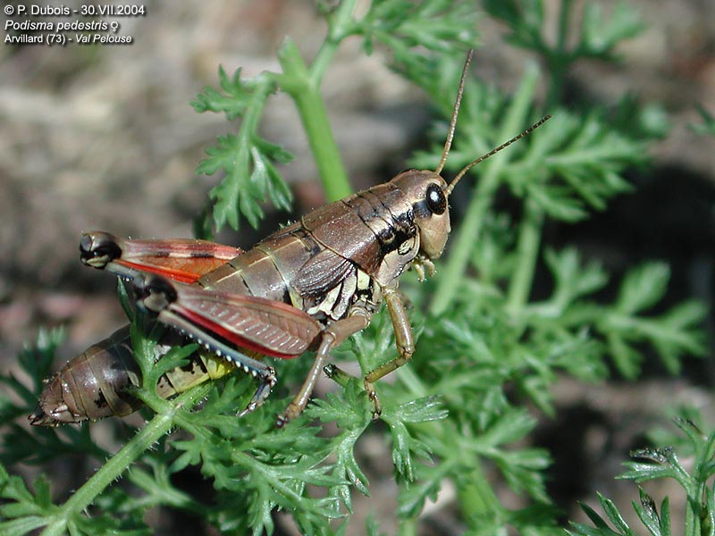
Does non-coding DNA have a function? Some of it does, to be sure. Some of it is involved in chromosome structure and cell division (e.g., telomeres, centromeres). Some of it is undoubtedly regulatory in nature. Some of it is involved in alternative splicing (Kondrashov et al. 2003). A fair portion of it in various genomes shows signs of being evolutionarily conserved, which may imply function (Bejerano et al. 2004; Andolfatto 2005; Kondrashov 2005; Woolfe et al. 2005; Halligan and Keightley 2006). On the other hand, the largest fraction is comprised of transposable elements — some of which become co-opted by the host genome, some of which play major role in generating genomic variation, some of which may be involved in cellular stress response, and yet others of which remain detrimental to host fitness (Kidwell and Lisch 2001; Biémont and Vieira 2006). The upshot is that some non-coding DNA is most certainly functional — but when it is, this usually makes sense only in an evolutionary context, particularly through processes like co-option. More broadly, those who would attribute a universal function for non-coding DNA must bear the following in mind: any proposed function for all non-coding DNA must explain why an onion or a grasshopper needs five times more of it than anyone reading this sentence.
Should “junk” be thrown out?

There is nothing wrong with a word taking on a new meaning as knowledge changes – that is, unless reference to an original (and outmoded) sense lingers as a source of confusion, or the term expands so much as to lose contact with an initially accurate definition. Indeed, even the term “evolution” is technically a misnomer since its etymology implies an “unfolding”, as of a pre-determined developmental program (see Bowler 1975). The objection raised here is not to terms that change in usage per se, but to those whose shifting usage involves collecting or retaining unwanted conceptual baggage. This is especially relevant when the baggage is toted surreptitiously (note that no serious biologist takes “evolution” to mean a pre-determined unfolding but that ideas of inherent “progress” have been almost impossible to shake; see Gould 1996; Ruse 1996).

“Junk DNA”, which originally was coined in reference to now-functionless gene duplicates (i.e., true broken-down “junk”), is now used as “a catch-all phrase for chromosomal sequences with no apparent function” (Moore 1996). Its current usage also implies a lack of function which is accurate by definition for pseudogenes in regard to protein-coding, but which does not hold for all non-coding elements. The term has deviated from or outgrown its original use, and its continued invocation is non-neutral in its expression – and generation – of conceptual biases.

“Junk DNA” is not the only offender. Non-coding DNA has been called by many names that have had the same pejorative undertones (intentional or not) implying uselessness, if not outright wastefulness. Examples include excess DNA (Zuckerkandl 1976; Doolittle and Sapienza 1980), surplus or nonessential or degenerate or silent DNA (Comings 1972; Gilbert 1978), quiet DNA (Lefevre 1971), garbage DNA (Ohno 1970), non-informational or nonsense DNA (Ohno 1972b), worthless DNA (Ohno 1973), trivial DNA (Ohno 1974), vestigial DNA (Loomis 1973), redundant DNA (Vinogradov 1998), supplementary DNA (Hutchinson et al. 1980), secondary DNA (Hinegardner 1976), and incidental DNA (Jain 1980).

As Gould (2002, p.503) stated, “A rose may retain its fragrance under all vicissitudes of human taxonomy, but never doubt the power of a name to shape and direct our thoughts”. Because it is generally no longer applied in its original meaningful sense, because the type of DNA to which it actually relates now has a more descriptive name (pseudogenes), and because of its connotations of total phenotypic inertness, the term “junk DNA” should probably be abandoned in favour of less subjective terminology. “Non-coding DNA” serves this purpose quite well.

Concluding remarks

It is an exciting time in genome biology. Aspects of genomic form and function that were largely inconceivable only a few decades ago are now being revealed on a daily basis. It should come as no surprise (and indeed, it probably does not) that new roles are being discovered for non-coding DNA and that some of yesterday’s buzzwords — including “junk DNA” — are destined for the dustbin. However, extrapolating each report that a given small segment of DNA may be functional to mean that all non-coding DNA is vital is as counterproductive as dismissing non-coding DNA as totally non-functional. Genomes are complex, and there is little use in approaching them from a simplistic point of view.

——

Andolfatto, P. 2005. Adaptive evolution of non-coding DNA in Drosophila. Nature 437: 1149-1152.

Batten, D. 1998. ‘Junk’ DNA (again). Creation Ex Nihilo Technical Journal 12: 5.

Beaton, M.J. and T. Cavalier-Smith. 1999. Eukaryotic non-coding DNA is functional: evidence from the differential scaling of cryptomonad genomes. Proceedings of the Royal Society of London, Series B 266: 2053-2059.

Bejerano, G., M. Pheasant, I. Makunin, S. Stephen, W.J. Kent, J.S. Mattick, and D. Haussler. 2004. Ultraconserved elements in the human genome. Science 304: 1321-1325.

Bennett, M.D. 1982. Nucleotypic basis of the spatial ordering of chromosomes in eukaryotes and the implications of the order for genome evolution and phenotypic variation. In Genome Evolution (eds. G.A. Dover and R.B. Flavell), pp. 239-261. Academic Press, New York.

Bergman, J. 2001. The functions of introns: from junk DNA to designed DNA. Perspectives on Science and Christian Faith 53: 170-178.

Biémont, C. and C. Vieira. 2006. Junk DNA as an evolutionary force. Nature 443: 521-524.

Bostock, C. 1971. Repetitious DNA. Advances in Cell Biology 2: 153-223.

Bowler, P.J. 1975. The changing meaning of “evolution”. Journal of the History of Ideas 36: 95-114.

Bremmerman, H.J. 1987. The adaptive significance of sexuality. In The Evolution of Sex and its Consequences (ed. S.C. Stearns), pp. 135-161. Birkhauser Verlag, Basel.

Brenner, S. 1998. Refuge of spandrels. Current Biology 8: R669.

Britten, R.J. and E.H. Davidson. 1969. Gene regulation for higher cells: a theory. Science 165: 349-357.

Britten, R.J. and E.H. Davidson. 1971. Repetitive and non-repetitive DNA sequences and a speculation on the origins of evolutionary novelty. Quarterly Review of Biology 46: 111-138.

Castillo-Davis, C.I. 2005. The evolution of noncoding DNA: how much junk, how much func? Trends in Genetics 21: 533-536.

Comeron, J.M. 2001. What controls the length of noncoding DNA? Current Opinion in Genetics & Development 11: 652-659.

Comings, D.E. 1972. The structure and function of chromatin. Advances in Human Genetics 3: 237-431.

Dawkins, R. 1999. The “information challenge”: how evolution increases information in the genome. Skeptic 7: 64-69.

Doolittle, W.F. and C. Sapienza. 1980. Selfish genes, the phenotype paradigm and genome evolution. Nature 284: 601-603.

Doolittle, W.F. 1982. Selfish DNA after fourteen months. In Genome Evolution (eds. G.A. Dover and R.B. Flavell), pp. 3-28. Academic Press, New York.

Gall, J.G. 1981. Chromosome structure and the C-value paradox. Journal of Cell Biology 91: 3s-14s.

Georgiev, G.P. 1969. On the structural organization of operon and the regulation of RNA synthesis in animal cells. Journal of Theoretical Biology 25: 473-490.

Gibbs, W.W. 2003. The unseen genome: gems among the junk. Scientific American 289(5): 46-53.

Gibson, L.J. 1994. Pseudogenes and origins. Origins 21: 91-108.

Gilbert, W. 1978. Why genes in pieces? Nature 271: 501.

Gould, S.J. 1996. Full House. Harmony Books, New York.

Gould, S.J. 2002. The Structure of Evolutionary Theory. Harvard University Press, Cambridge, MA.

Halligan, D.L. and P.D. Keightley. 2006. Ubiquitous selective constraints in the Drosophila genome revealed by a genome-wide interspecies comparison. Genome Research 16: 875-884.

Hinegardner, R. 1976. Evolution of genome size. In Molecular Evolution (ed. F.J. Ayala), pp. 179-199. Sinauer Associates, Inc., Sunderland.

Hutchinson, J., R.K.J. Narayan, and H. Rees. 1980. Constraints upon the composition of supplementary DNA. Chromosoma 78: 137-145.

Jacq, C., J.R. Miller, and G.G. Brownlee. 1977. A pseudogene structure in 5S DNA of Xenopus laevis. Cell 12: 109-120.

Jain, H.K. 1980. Incidental DNA. Nature 288: 647-648.

Jerlström, P. 2000. Pseudogenes: are they non-functional? Creation Ex Nihilo Technical Journal 14: 15.

Kidwell, M.G. and D.R. Lisch. 2001. Transposable elements, parasitic DNA, and genome evolution. Evolution 55: 1-24.

Kondrashov, F.A. and E.V. Koonin. 2003. Evolution of alternative splicing: deletions, insertions and origin of functional parts of proteins from intron sequences. Trends in Genetics 19: 115-119.

Kondrashov, A.S. 2005. Fruitfly genome is not junk. Nature 437: 1106.

Lefevre, G. 1971. Salivary chromosome bands and the frequency of crossing over in Drosophila melanogaster. Genetics 67: 497-513.

Loomis, W.F. 1973. Vestigial DNA? Developmental Biology 30: F3-F4.

Makalowski, W. 2003. Not junk after all. Science 300: 1246-1247.

Max, E.E. 2002. Plagiarized errors and molecular genetics: another argument in the evolution-creation controversy. Talk.Origins Archive.

Moore, M.J. 1996. When the junk isn’t junk. Nature 379: 402-403.

Nowak, R. 1994. Mining treasures from ‘junk DNA’. Science 263: 608-610.

Ohno, S. 1970a. Evolution by Gene Duplication. Springer-Verlag, New York.

Ohno, S. 1970b. The enormous diversity in genome sizes of fish as a reflection of nature’s extensive experiments with gene duplication. Transactions of the American Fisheries Society 1970: 120-130.

Ohno, S. 1972. So much “junk” DNA in our genome. In Evolution of Genetic Systems (ed. H.H. Smith), pp. 366-370. Gordon and Breach, New York.

Ohno, S. 1973. Evolutional reason for having so much junk DNA. In Modern Aspects of Cytogenetics: Constitutive Heterochromatin in Man (ed. R.A. Pfeiffer), pp. 169-173. F.K. Schattauer Verlag, Stuttgart, Germany.

Ohno, S. 1974. Chordata 1: protochordata, cyclostomata, and pisces. In Animal Cytogenetics, Vol. 4 (ed. B. John), pp. 1-92. Gebrüder Borntraeger, Berlin.

Ohno, S. 1982. The common ancestry of genes and spacers in the euchromatic region: omnis ordinis hereditarium a ordinis priscum minutum. Cytogenetics and Cell Genetics 34: 102-111.

Ohno, S. 1985. Dispensable genes. Trends in Genetics 1: 160-164.

Patrushev, L.I. and I.G. Minkevich. 2006. Eukaryotic noncoding DNA sequences provide genes with an additional protection against chemical mutagens. Russian Journal of Bioorganic Chemistry 32: 1068-1620.

Petsko, G.A. 2003. Funky, not junky. Genome Biology 4: 104.

Raup, D.M. 1991. Exctinction. W.W. Norton & Co., New York.

Roels, H. 1966. “Metabolic” DNA: a cytochemical study. International Review of Cytology 19: 1-34.

Ruse, M. 1996. Monad to Man. Harvard University Press, Cambridge, MA.

Shapiro, J.A. and R. von Sternberg. 2005. Why repetitive DNA is essential to genome function. Biological Reviews 80: 227-250.

Sharma, A.K. 1985. Chromosome architecture and additional elements. In Advances in Chromosome and Cell Genetics (eds. A.K. Sharma and A. Sharma), pp. 285-293. Oxford and IBH Publishing Co., New Delhi.

Slack, F.J. 2006. Regulatory RNAs and the demise of ‘junk’ DNA. Genome Biology 7: 328.

Vinogradov, A.E. 1998. Buffering: a possible passive-homeostasis role for redundant DNA. Journal of Theoretical Biology 193: 197-199.

Walker, P.M.B., W.G. Flamm, and A. McLaren. 1969. Highly repetitive DNA in rodents. In Handbook of Molecular Cytology (ed. A. Lima-de-Faria), pp. 52-66. North-Holland Publishing Co., Amsterdam.

Walkup, L.K. 2000. Junk DNA: evolutionary discards or God’s tools? Creation Ex Nihilo Technical Journal 14: 18-30.

Wickelgren, I. 2003. Spinning junk into gold. Science 300: 1646-1649.

Wieland, C. 1994. Junk moves up in the world. Creation Ex Nihilo Technical Journal 8: 125.

Woodmorappe, J. 2000. Are pseudogenes ‘shared mistakes’ between primate genomes? Creation Ex Nihilo Technical Journal 14: 55-71.

Woolfe, A., M. Goodson, D.K. Goode, P. Snell, G.K. McEwen, T. Vavouri, S.F. Smith, P. North, H. Callaway, K. Kelly, K. Walter, I. Abnizova, W. Gilks, Y.J.K. Edwards, J.E. Cooke, and G. Elgar. 2005. Highly conserved non-coding sequences are associated with vertebrate development. PLoS Biology 3: e7.

Yunis, J.J. and W.G. Yasmineh. 1971. Heterochromatin, satellite DNA, and cell function. Science174: 1200-1209.

Zuckerkandl, E. 1976. Gene control in eukaryotes and the C-value paradox: “Excess” DNA as an impediment to transcription of coding sequences. Journal of Molecular Evolution 9: 73-104.

Zuckerkandl, E. and W. Hennig. 1995. Tracking heterochromatin. Chromosoma 104: 75-83.

Zuckerkandl, E. 1997. Junk DNA and sectorial gene expression. Gene 205: 323-343.

__________

Update: At Sandwalk, Larry Moran argues that the term “junk DNA” is “a good term”, “an accurate term”, and “a useful term”. You can read my response in the comments section of the original post or in my re-post on this blog.